Chapter Four—
A Comparative Study of the Levels and the Differentials of Sterility in Cameroon, Kenya, and Sudan
Ulla Larsen
Children are the cloth of the body.
Without children you are naked.
Yoruba saying (Nigeria)
Numerous populations in sub-Saharan Africa experience very low fertility (Page and Coale, 1972). Effective intentional practices to lower reproduction are uncommon in Africa and most populations approximate natural fertility. The occurence of infertility is a major problem for individuals, since most Africans desire large families (at least six children). Low fertility also indicates serious health problems, because low fertility is strongly linked to sexually transmitted diseases, such as gonorrhea. If the prevalence and incidence of sterility were better understood, more effective campaigns to reduce sterility could be carried out. However, if the levels of sterility were reduced, fertility and the population growth rate would most likely increase rapidly, thereby causing other problems. Thus, the prevalence of sterility in certain areas of sub-Saharan Africa and the effective prevention or treatment of sterility are complex issues.
Sterility is defined as the inability of a noncontracepting, nonlactating, sexually active woman to have a live birth. Primary sterility is defined biologically as never developing the capacity to reproduce, whereas secondary sterility refers to the termination of previously possessed reproductive ability. In practice, sterility is difficult to measure. Celibate women are never at risk of having a child or of testing their reproductive capacity. Therefore, in establishing operational definitions, only noncelibate women are considered. Those who have never had a live birth are usually considered to have primary sterility, while secondary sterility is measured among women who are unable to have a live birth subsequent to an earlier live birth.
Studies of sterility in Africa have been hampered by the fact that no method of measuring the levels of sterility by age from incomplete birth histories has been available until now (Larsen, 1985). In addition, until recently demographic data for sub-Saharan Africa have been both scarce and of poor
quality. In the last decade several sources have become available; surveys, carried out by several African countries in collaboration with the World Fertility Surveys (WFS), provide a particularly rich body of information. The present study is based on WFS data from Cameroon, Kenya,[1] and Sudan.[2] These three countries were selected because they represent many of the variations in reproductive characteristics in sub-Saharan Africa, such as difference in age at first marriage and duration of postpartum abstinence (Lesthaeghe, 1984b). Total fertility is relatively low in Cameroon and Sudan, and relatively high in Kenya; Sudan is the only country for which the WFS included questions about female circumcision.
In this chapter, the discussion begins with a description of the geographic distribution of subfertility in sub-Saharan Africa. Next, theoretical models of sterility are established, previous research on differentials in subfertility is reviewed, and the covariates of sterility to be examined in this study are defined. Subsequently, age-specific sterility rates and the levels of primary sterility are estimated for all women and for selected subgroups. Finally, in order to examine the effects of several covariates simultaneously on the prevalence or incidence of sterility at different time points or across cohorts, a hazards models analysis is conducted.
Subfertility in Sub-Saharan Africa
Sterility is strongly associated with low levels of fertility. Therefore, a map of the geographic variations in the levels of fertility for peoples of Africa south of the Sahara provides a good indication of the prevalence of sterility. Page and Coale (1972) estimated total fertility rates for all the populations in sub-Saharan Africa for which data were available (figure 4.1). Their study shows that the low fertility areas are concentrated in Central Africa and include, among other countries, Cameroon, Central African Republic, the western part of Sudan, Congo, and Gabon. Recently, this distribution pattern of sub-fertility has been confirmed by Frank (1983a , 1983b ), who mapped the proportion childless among women aged 45–49.
Models of Sterility
A couple's inability to have a live birth may be due to impairment of the reproductive system of the wife, the husband, or both. In general, female factors are thought to cause sterility in 50 to 70 percent of all sterile couples (Sherris and Fox, 1983). However, the contribution of female factors might be overestimated, since sterility investigations traditionally have concentrated on women rather than men.
Female sterility is primarily due to one or more of the following reasons: (1) ovaries may fail to produce a viable egg, preventing the possibility of
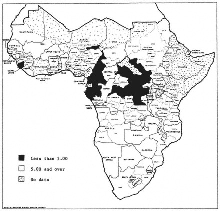
Figure 4.1.
Total Fertility Rates for Selected Areas or Peoples of Africa
South of the Sahara (Source: Page and Coale, 1972)
conception; (2) fallopian tubes may be blocked, distorted, or infected, preventing normal movement of the egg or sperm in the tubes; (3) the uterus may be distorted or the uterine lining (endometrium) inadequate or infected, preventing implantation or survival of the embryo; (4) the cervix may be malformed, infected, or secrete abnormal mucus, preventing sperm from reaching the upper reproductive tract; and (5) systemic infection or hormonal imbalance may result in fetal death (figure 4.2).
Sterility is most frequently caused by a pelvic inflammatory disease (PID), which originates in the cervix and can ascend to the upper reproductive tract and block the fallopian tubes. PID can also lead to fluid filled swellings, adhesions, scarring, and other permanent damage of the fallopian tubes. Permanent sterility is more likely to occur if the infection is severe, if treatment is delayed, or if a woman has had multiple episodes of PID. An
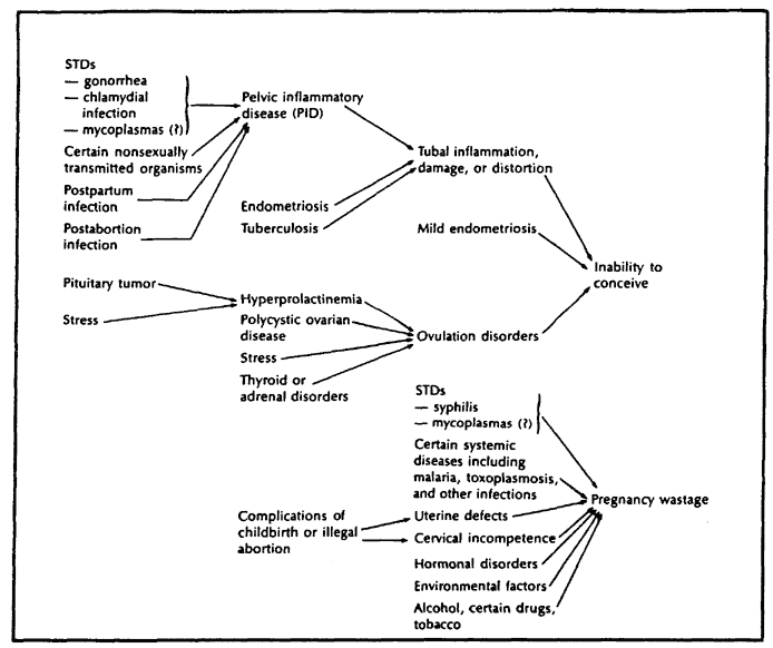
Figure 4.2.
Relationships of Selected Direct and Indirect Causes of Female Infertility
(Source: Sherris and Fox, 1983)
estimated 60 percent of women with PID become sterile if they are not treated with antibiotics (Sherris and Fox, 1983). The most common source of PID is a sexually transmitted disease, such as gonorrhea and chlamydia. However, the risk of PID is also fairly high when childbirth or abortion is carried out under unhygienic conditions, because microorganisms ascend more easily through a dilated cervix.
Sterility can result from the inability to carry a pregnancy to term as well as the inability to conceive. Pregnancy wastage often results from causes that cannot be prevented, such as abnormalities of the fetus and problems with the uterus or cervix. Other causes are preventable and they include sexually transmitted diseases, such as syphilis, and certain systemic diseases, such as malaria. The risk of pregnancy wastage is high for women with syphilis because this disease can infect the fetus, causing intrauterine death. Malaria can result in pregnancy wastage because malarial infection of the placenta impairs fetal nutrition and increases the risk of spontaneous abortion.
Female circumcision has been indicated as increasing the risk of sterility,
but evidence is lacking. It is hypothesized that circumcision predisposes women to infections leading to sterility because scarring and closure of the external genitalia prevent proper drainage of urine and menstrual blood. Also, circumcised women often experience pain during sexual intercourse and difficulties at childbirth. All of these problems may lead to primary sterility. In addition, the risk of secondary sterility may be increased because at each delivery most circumcised women have to be cut to allow delivery of the fetus and the incision is subsequently stitched. This procedure frequently causes infections and subsequent sexual intercourse is often more difficult, even impossible in some cases. It should be noted that there are different types of female circumcision. The most common is a Pharaonic circumcision, which is a removal of the clitoris, the labia minora, the labia majora, and a closing of the vagina to only a small opening to allow elimination of urine and menstrual blood. Less common is a Sunna circumcision, where only the prepuce of the clitoris is removed. Other types are also performed which vary in degree between a Sunna and a Pharaonic circumcision. It is generally agreed that a Pharaonic circumcision may especially enhance the risk of sterility (Aziz, 1980; People, 1979; Shandall, 1967).
Male sterility is usually due to blockage of sperm ducts, or disorders in sperm production resulting in poor semen quality, that is, semen that contains too few sperm and/or abnormal sperm (figure 4.3). A less common cause is a sexual malfunction that prevents ejaculation of semen. Genital
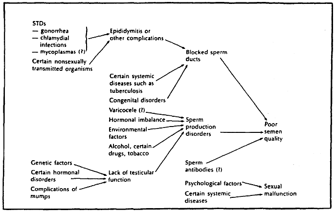
Figure 4.3.
Relationships of Selected Direct and Indirect Causes of Male Infertility
(Source: Sherris and Fox, 1983)
infections cause poor semen quality and sterility by creating inflammation or blockage in the upper reproductive tract. The infection is usually due to sexually transmitted diseases, such as gonorrhea and chlamydia, but it can also be caused by urogenital tuberculosis and other diseases (Sherris and Fox, 1983).
The Covariates of Sterility
Empirical analyses of sterility in sub-Saharan Africa are complicated by the fact that data about the major factors directly related to sterility are usually not available. Therefore, most previous research on sterility in sub-Saharan Africa has used indirect evidence of sterility, and variables related only indirectly to sterility, as does this study. The variables included are selected on theoretical grounds and on the basis of the literature on factors related to subfertility. Clearly these factors must be related to be health problems described in the previous section, although our analysis cannot delineate these relationships because of the lack of available data.
The major limitations of the WFS surveys as a source of information about sterility are as follows. First, the WFS surveys provide only data about women's reproductive histories; hence, sterility of a couple is ascribed always to the woman. Second, no data are available about women's medical histories, for example, whether a woman has had a venereal disease or whether she has had medical treatment. Third, the WFS surveys collected no information about location of childbirth and about who assisted with the delivery (midwifery) for Kenya or Sudan. In Cameroon, midwifery information is provided only for the last pregnancy. Finally, the WFS surveys asked questions about female circumcision only in Sudan.
As a consequence of the lack of data about medical history, midwifery, and female circumcision, proxies are used for health information. One further problem with these data must be noted. There is evidence indicating that the prevalence of venereal diseases leading to sterility has decreased since knowledge about the effects of antibiotics became available around 1950 (Tabutin, 1982). Infections following a childbirth or an abortion can also be cured with penicillin, and midwifery is likely to have improved in the last few decades. Furthermore, in Sudan the ratio of Sunna to Pharaonic circumcisions has increased slightly, suggesting that complications leading to sterility from female circumcision may have decreased (Shandall, 1967). Thus, it is possible that place of the delivery, type of attendant at the birth, and circumcision have different relations to sterility in different cohorts, that the levels of sterility by age have declined in recent decades, and that the prevalence of sterility may vary across cohorts.
The WFS surveys generally provide information about socioeconomic variables only at the time of survey, so that we are forced to limit the analysis
to a time period during which it is reasonable to assume that the covariates do not change. Due to this restriction, two models are established: model 1 of sterility status (sterile or fecund) 5 years before survey (we need at least 5 years of exposure to determine sterility status) includes socioeconomic variables; model 2 of sterility status at different points in time or by cohorts excludes most of these variables. In model 1 it is assumed that the single observation made on a covariate (such as education) at the time of the survey holds for the previous 5 years. This assumption should not bias the results substantially because none of the socioeconomic variables analyzed change much in 5 years. For instance, in the societies studied, few married women move or start to go to school and most husbands do not change their work statuses. However, it is not valid to assume that all covariates are constant from entry into first marriage until survey; for example some women divorce and other women acquire cowives. Therefore, model 2, which looks at women at 5-year age intervals over their entire married lifetime, is restricted to either constant covariates or to time-varying covariates for which data at different points in time are available.
Both models include the following as covariates: region, religion, residence (urban or rural), education, type of circumcision, number of times married, use of contraception, and age. Model 1 adds the covariates: husband's education, husband's work status, and marital status (monogamous versus polygamous unions and wife order). Model 2 includes the basic set of covariates plus time period and cohort. The justification for inclusion of these variables is described below.
Geographic variations in sterility can provide clues to the presence of environmental variations (such as prevalence of venereal diseases) related to sterility, and to cultural patterns that affect sexual practices (such as more sexual partners). Subfertility has been found to vary greatly from region to region in all three countries. For instance, the percent childless is higher in the North, Center-South and East regions than in the West regions of Cameroon (Santow and Bioumla, 1984; Nasah, Azefor, and Ondoa, 1974). Furthermore, previous research shows that geographic boundaries of subfertility are often ethnic boundaries (Caldwell and Caldwell, 1983; Retel-Laurentin, 1974; Romaniuk, 1968). For example, the Mijikendas and the Kikuyus are neighboring tribes in Kenya and the former group has much lower fertility than the latter (Kenya Fertility Survey 1977–1978, 1980). Lesthaeghe (1985) and Retel-Laurentin (1974) noted that in subfertile societies, marital mobility is high and tribal customs permit wider sexual contacts, while ethnic groups that practice strict marriage laws have higher fertility.
Region is used as a proxy for venereal diseases and the sexual practices associated with its frequency. It would be preferable to use ethnicity instead of region, but no data are available about ethnic groups in Sudan. There are also too many distinct ethnic groups in Cameroon (thirty-four) and Kenya
(forty-three) to examine the levels of sterility across ethnic groups. In a previous study of Cameroon and Kenya, ethnic groups located in the same area were combined and it was found that the differentials in sterility by ethnicity are very similar to the variations across region (Larsen, 1985). It is problematic, however, to use region in model 2, due to the fact that some women migrate after first marriage. More specifically, if some women move from a region with low sterility to a region with high sterility, then the estimated effects for the latter region at survey will be biased downwards, indicating lower sterility than is the case, because some women actually spent a part of their life in a more healthy environment. Likewise, migration in the opposite direction will cause the effects estimates for the low sterility region to be biased upwards, masking the explanatory power of region.
Romaniuk (1968) pointed out that low fertility areas often are close to waterways, such as lakes, rivers, and the sea, and that subfertility seems to follow the Arab slave routes. In accordance with this finding Romaniuk observed that Muslims suffer more from subfertility than non-Muslims. Today, it is uncertain whether the Arab slave traders spread venereal diseases, thus causing sterility, or whether the prevalence of sterility in these areas is predominantly due to cultural practices among Muslims. For instance, female circumcision is more frequently practiced among Muslims than non-Muslims, although female circumcision is not an ordinance from Islam (Shandall, 1967). Religion is included for Cameroon and Kenya, but not for Sudan, where only 38 women are non-Muslim in a sample of 3115 women (the WFS survey is restricted to northern Sudan). On the other hand, the effects of type of female circumcision are analyzed only for Sudan (no data about female circumcision are available for Cameroon and Kenya). The differentials between women with a Pharaonic circumcision and other women and examined (as few as 112 women (3.6 percent of the women surveyed) were not circumcised, so the differentials between never circumcised and circumcised women cannot be analyzed).
Urban or rural residence is hypothesized to be linked to the prevalence of venereal diseases and different practices of midwifery. In general, prostitution is more common in cities; hence, exposure to venereal diseases and subsequent sterility is likely to be higher in urban than in rural areas, even though medical treatment is more readily accessible in urban areas. Conversely, secondary sterility due to infections from a previous childbirth or abortion is probably greater for women who live in rural areas where more births are delivered at home under poor sanitary conditions by a family member or an untrained midwife. Previous research shows that the pattern of low fertility by residence is not systematic; subfertility is more widespread in rural than in urban areas in Central Africa, while the Yorubas in Nigeria experience very little difference in fertility by residence, and fertility is higher in rural than urban Ghana (Caldwell and Caldwell, 1983). In order to deter-
mine the impact of residence on sterility and to test the hypothesis that barren women tend to migrate from rural to urban areas to hide their childlessness (Frank, 1983b ), the differentials in sterility by residence and whether a woman has migrated since her childhood (moved from a rural to an urban area), are analyzed.
Education of the wife and of her husband and husband's work status are used as indicators of socioeconomic status. Education is assumed to be linked to lower sterility because educated people are better informed. As a consequence, they are more likely to seek treatment for a venereal disease, and they are probably better able to avoid an infection leading to sterility, for example, by being more aware of the importance of hygienic conditions at childbirth. Education is also generally associated with higher income, and educated people can therefore better afford medical treatment and sanitary facilities. Information about education is available only at survey. It is assumed that women did not continue their education after they married, so this variable is included in both models. Education of the husband and his work status are included only in model 1 because some women divorce and do not have the same husband from the time of their first marriage until the survey. It is hypothesized that the employers (men who employ one or more people) have the highest income and are the best informed, that men who are self-employed (for example, in agriculture or trade) are more traditional and poorer, while the employees are a heterogeneous group.
In many African societies it is considered the husband's right to have offspring, and childlessness or subfertility is usually blamed on the wife. It is common practice to dissolve subfertile unions; consequently, as a group, divorced women have lower fertility. However, most divorced women remarry shortly thereafter and often become higher order wives in polygamous unions. Not surprisingly, polygamous unions generally have lower fertility than monogamous unions (Frank, 1983b ; Henin, 1981). A study of Cameroon and Ghana shows that the proportion of women with no live birth at any expected duration of marriage is lower for women of rank 1 in polygamous unions compared to women in monogamous unions, while women of rank 2 and rank 3+ in polygamous unions have the highest proportion of childlessness (Lesthaeghe, 1984a ). For other regions, subfertility might be higher in polygamous than monogamous unions because a man adds a new wife to a subfertile union to achieve higher parity (Henin, 1981).
To test whether women married more than once have higher sterility, the covariate, number of times married, is included in both model 1 and model 2. In model 2, number of times married may, of course, change with age but the marital history of each woman in the WFS surveys provides this information. The effects of marital status are examined only in model 1 because the dates cowives entered polygamous unions are not available. It is, therefore, assumed that marital status did not change in the 5 years preceding the
survey. Marital status is grouped differently in the three countries analyzed because the individual WFS questionnaires varied; for example, the survey for Sudan provides no data about wife order in polygamous unions.
In order to measure the effects of contraception on sterility, it would be ideal to know the ages at which each woman starts and stops using contraception. However, the WFS surveys only provide data about whether a woman has ever used and whether she is currently using contraceptives at survey. Women who have used efficient methods of contraception during a long period have lower fertility and are very likely falsely to be classified as sterile. Many women in the societies studied do not contracept efficiently, and it is hypothesized that contraception only decreases fecundity slightly. In contrast, some methods of contraception (such as the condom and diaphragm) protect against venereal diseases and subsequent sterility. In addition, women who have used contraception tend to have more education and are probably more inclined to seek treatment for venereal diseases and to have trained assistance at childbirth, two factors that protect against sterility. Information about the ever use of contraception is employed in the present study, because it covers both past and current users. We are forced to assume that women who report that they have used contraception were contracepting throughout their entire married life, although most of these women probably contracepted only in certain periods. Consequently, the direct effects of contraception on fecundity are measured very poorly, while the indirect effects of being better informed probably vary less with time.
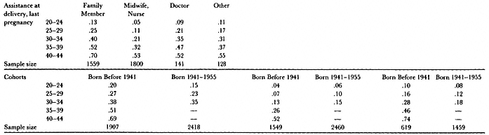
In Cameroon, information about midwifery (location at delivery and assistance of delivery) is available for the last pregnancy, as mentioned previously. However, it is problematic to interpret the effects of midwifery on sterility in analyses of individual women. For instance, women who delivered their last birth in a hospital or who were assisted by a doctor may have higher sterility than other women, due to the fact that these women experienced complications at their last delivery and therefore received special medical care. Otherwise, we would assume that women who delivered their last birth in a hospital or were assisted by a doctor are less likely to be sterile than women who delivered at home or were assisted by an untrained midwife. Furthermore, since data about midwifery are available only about the last pregnancy; it is not possible to determine the effects of midwifery on sterility throughout a woman's reproductive life, since we would be forced to make the false assumption that midwifery had been the same for all births. Due to these shortcomings, the present study examines the effects of midwifery on sterility only at the group level.
There are three major dependent variables: levels of primary sterility, incidence of secondary sterility, and prevalence of sterility (both primary and secondary). These variables are measured both at the group and at the individual level. For example, the prevalence of sterility by age group in a
population or one of its subgroups is estimated at the group level without assigning an age at sterility to individual women in that group. At the individual level, each woman must be assigned a sterility status at each age. Since the second procedure is far more subject to error, both are used in different circumstances. Depending on the question to be addressed, the age range of women included in a particular analysis varies. For estimates of levels of primary sterility, only women married before age 20 are considered. (It becomes very difficult to distinguish between biological primary sterility and early secondary sterility in populations where menarche precedes the initiation of sexual relations by a relatively long time.) At the group level, age-specific prevalence rates of sterility are estimated by standard 5-year age intervals for women in the age range from 20–45. Sterility below age 20 is not examined in these analyses because of the complications caused by adolescent subfecundity. For women above age 45 the ability to have a live birth decreases sharply and biological aging is the predominant cause of sterility. At the later ages of the reproductive span, it also becomes more difficult to distinguish sterile from fecund women, a problem that is especially pronounced in studies of individual women (Larsen, 1985). Therefore, at the individual level, the analysis of the covariates of the incidence of sterility is limited to the age range from 20–40, while the covariates of the prevalence of sterility are analyzed for the age interval 20–45.
Finally, in order to determine a woman's status (sterile or fecund) at any age, at least 5 subsequent years of exposure are needed (the methods of measuring sterility are described below). Consequently, the closest to interview we can determine current sterility status is 5 years before survey, as utilized in model 1. In model 2 each woman's status is determined 5 years before survey, 10 years before survey, and so forth. The oldest women entered first marriage so long ago that we can determine their status 25 years before survey. As an alternative variable to time period, the link between different cohorts and sterility may be analyzed. In model 2 the cohorts examined are women born in the following periods: before 1933, 1933–1937, 1938–1942, 1943–1947, and 1948 and later.
Methods of Measuring Sterility
Primary sterility is measured among women who married below age 20 by the proportion childless after at least 7 years of marriage (Larsen, 1985; Vincent, 1950). Age-specific prevalence rates of sterility are estimated by a measure originally proposed by Louis Henry (1965, 1961) and termed "the proportion subsequently infertile." This method substitutes the number of infertile couples at a given age for the number of sterile couples at that age, where a couple is defined as being infertile if it has no live birth at that age or later (at least the next 5 years, if incomplete birth histories are used). The proportion sterile is then estimated as the number of infertile couples at a
given age divided by all couples at that age. The relation between the estimated proportion sterile at a given age and the age at which this proportion sterile is attained was determined in a simulation study where the exact age at sterility of each woman as well as the exact proportion sterile at each age is known (Larsen, 1985).
An individual woman's status (sterile or fecund) at particular ages is determined on the basis of the length of the open birth interval. Women whose last live birth occurred less than 5 years before censoring are assigned the status of fecund when last observed; otherwise an age at onset of sterility is assigned. The age assigned is determined by the age at the last live birth (or marriage, if childless), but it is generally not equal to the age at the last reproductive event; the latter age usually precedes the actual age at onset of sterility. The correspondence between an individual woman's age at the last reproductive event and the age at onset of sterility has been determined in a simulation study (Larsen, 1985).
The introduced group and individual measures of sterility are robust to interpopulation variations in reproductive characteristics (age at marriage, the age pattern of sterility, and so forth) and to sampling variation in samples of at least 500 women. The primary sterility estimate requires a sample of at least 1000 women. Finally, individual assignments of ages at onset of sterility are quite sensitive and specific, although the accuracy declines with age (Larsen, 1985).
The Levels of Sterility in Cameroon, Kenya, and Sudan
This section examines the prevalence of primary sterility and the levels of sterility by age for all women in a population and for selected subgroups. In order to carry out this analysis we need to define the sample of women who have been exposed to childbirth.
Only women in the age range from 15 to 50 who have been in one union for at least 5 years are considered. The required 5-year period of exposure might introduce a bias, because subfertile unions are frequently dissolved and even though divorced women usually remarry, they may not have been in a union 5 years or longer at censoring. The period of exposure is assumed to begin at first marriage, but there is evidence indicating that women are often sexually active prior to marriage (see Lesthaeghe, 1984a ). This bias has little effect on the results, since the group measures of sterility and the assignment of individual ages at onset of sterility are quite robust to misassignment of age at first marriage (initiation of sexual relationships) (Larsen, 1985). Exposure ends at the time of survey, if the first marriage is intact, or at the dissolution of first marriage. In the latter case, the woman reenters the analysis at entry into second marriage, and so forth.
The study of sterility is complicated by the fact that not all women in the
married state are sexually active. Some women practice postpartum abstinence, while other women have terminated all sexual activity, or are temporarily contracepting. Fortunately, the group and individual measures of sterility are not sensitive to variation in the period of postpartum abstinence (Larsen, 1985). The effects of terminal abstinence are also negligible, but only because it is rarely practiced. Only 2 percent (seventy-two women) in Cameroon and 1 percent (forty-eight women) in Kenya report terminal abstinence. No information is available about this question for Sudan.
Most people in Cameroon, Kenya, and Sudan want large families. Nevertheless, as many as 13 percent (564 women) in Cameroon, 32 percent (1306 women) in Kenya, and 24 percent (476 women) in Sudan report having used contraception. Consequently, the estimates of sterility may be biased, because fecund women who have contracepted efficiently over a long period of time might be falsely classified as sterile. Hence, to determine the effects of contraception on sterility, the levels of sterility by age are estimated separately for all women and for women who have never used contraception. Contraception appears to have almost no effect on the levels of sterility in these countries (table 4.1). In Sudan, only above age 35 is there slightly higher sterility for all women compared to women who have never used contraception. In Cameroon and Kenya women who have never used contraception have slightly higher age-specific rates of sterility than all women. These differences are small, so all women are included in the subsequent analysis.
The levels of sterility by age are highest in Cameroon, intermediate in
| ||||||||||||||||||||||||||||||||||||||||||||||||||||||||||||||||||||||||||||||||||||||||
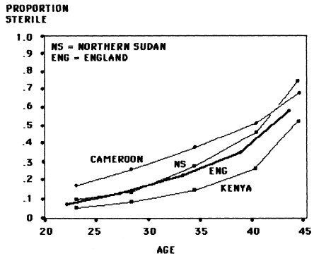
Figure 4.4.
Age-Specific Sterility Rates in Three African Countries
Compared to Those in Prenineteenth-Century England
Sudan, and lowest in Kenya for all ages below age 40 (figure 4.4). The age structure of the estimates is similar to that of a natural fertility population sample derived from reconstitutions of English families from 1550 to 1850 (Trussell and Wilson, 1985). The proportion sterile is systematically lower in Kenya than in England and systematically higher in Cameroon, while the proportion sterile in Sudan rises more steeply with age than in any of the other three populations. The levels of primary sterility for all women follow the same pattern as the age-specific rates of sterility (table 4.2). It is possible, however, that the estimates of primary sterility are too low because childlessness is probably underreported. There is also evidence of age misreporting
| ||||||||||||||||
and time misplacements of births and the impact of these reporting errors will be discussed later in this paper.
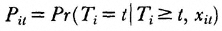
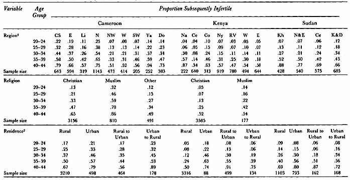
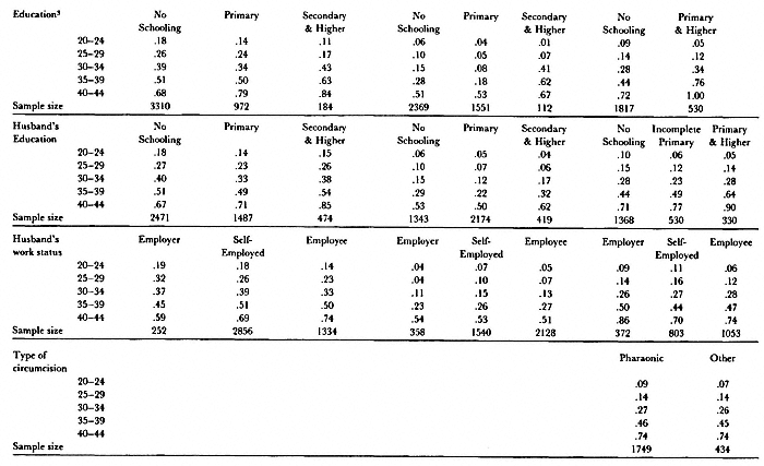
As a first step in the analysis of covariates of the prevalence of sterility, the samples of all women in Cameroon, Kenya, and Sudan were divided into subgroups defined by a single demographic or socioeconomic characteristic and the levels of sterility within groups was estimated. Variables had to be considered singly to maintain adequate sample sizes within each group. The characteristics analyzed include region, religion, residence, education, husband's education, husband's work status, type of circumcision, number of times married, marital status, location at delivery of last pregnancy, assistance of delivery of last pregnancy, and cohort.
The within-group differentials in sterility by demographic and socioeconomic factors are quite consistent across Cameroon, Kenya, and Sudan (tables 4.3 and 4.4). These observed differentials in sterility within a group may not be due to an association between the examined variable and sterility. Instead, they may be confounded by the effects of other covariates associated with sterility. For instance, uneducated women and women in certain regions appear to have higher sterility than educated women and women in other regions. However, it is possible that educated women live in regions with low sterility and that the apparent differentials by education are due to differences by region. In order to measure differentials in sterility within a variable and across variables it is necessary to estimate the effects of those variables hypothesized to be associated with sterility simultaneously. The requisite analysis cannot be carried out at the group level because of the sample size questions already described. Therefore, an individual level analysis was carried out to determine the effects of each variable simultaneously with several other variables hypothesized to be associated with sterility.[3]
Methods
In order to analyze simultaneously the effects of several demographic and socioeconomic factors on sterility at different points in time or by cohort, a hazards model approach is used. In general, a hazard or risk is the conditional probability of the occurrence of an event (dying, becoming sterile, and so forth) at a given age, provided that one has survived to that age without the event occurring. For example, assume that time can take only positive values (t = 1, 2, 3, . . .) and that we observe a total of n independent individuals (i = 1, . . ., n ) beginning at time t = I[4] . Then the discrete-time hazard rate is defined as
where T is a variable giving the time of event occurrence, and xit is a vector of explanatory variables, which may take on different values at different discrete times.
|
|
|
|
| ||||||||||||||||||||||||||||||||||||||||||||||||||||||||||||||||||||||||||||||||||||||||||||||||||||||||||||||||||||||||||||||||||||||||||||||||||||||||||||||||||||||||||||
| ||||||||||||||||||||||||||||||||||||||||||||||||||||||||||||
In the present study the logistic regression function is used to specify how the hazard rate depends on time and the explanatory variables. The explanatory variables for individuals i are in the form of a vector xit = (xi1t , xi2t , . . . , x ijt ), where there are j variables. Here, the value for an individual on a specific characteristic variable can vary over time. Then the logistic regression function for individual i at time t is
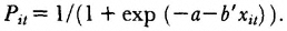
In logit form, this becomes
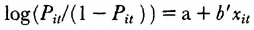
where a is a constant and b́ is the vector of regression coefficients associated with the risk factors xit . When the risk factors xit are restricted to be categorical variables, the regression coefficients can be used to determine the odds of the occurrence of the event under study, where the odds are defined as the ratio of the probability the event occurs to the probability it does not occur. In this case, all the xijt are dummy variables where xijt , equals 1 if individual i is in the category represented by the jth variable and is zero otherwise. The predicted odds of an event occurring for individual i in interval t are exp (a + b ́ xit ).
The ratio of two odds is called the relative odds. Hence, the odds at a given time for individual i relative to the odds for individual i ́ are exp (a + b ́ xit –a –b ́x ít ) In general, it is of interest to determine the relative odds when all factors but one are the same for the two groups of people. For example, if there is a three-category variable represented by two dummy
variables xi1t and xi2t , and individual i has xi1t = 1 and xi2t = 0, whereas individual i ́ has x í1t = 0 and x í2t = 1, then the relative odds (RO ) are exp (b1 – b2 ). Thus, for a given variable, if RO is greater than 1, then the odds of an event are higher for individual i ́ who is in category 1 relative to individual i' who is in category 2; if RO is less than or equal to 1, those in category 1 have lower than or the same odds of an event as those in category 2 when the categories on all other variables are exactly the same.
In practice, this procedure treats each discrete age interval for each individual as a separate observation or unit of analysis. For each of these observations in the incidence analysis, the dependent variable is coded 1 if an event occurred to that individual in that age interval; otherwise it is coded o. Thus, if an individual experienced an event at age interval 5, at least five different observations could be created. For the fifth observation the dependent variable would be coded 1. For the other four observations, the dependent variable would be coded o. In the prevalence analysis, the dependent variable is coded 1 if an event occurred in that age interval or in a previous age interval; otherwise it is coded 0.
The explanatory variables for each of these new observations would be assigned whatever values they had at that particular unit of age. When prevalence rates (such as, the probability of being sterile) are estimated, all individuals exit the analysis at censoring, whereas when incidence rates (for example, the probability of becoming sterile) are estimated, individuals exit the analysis either at censoring or at the time of the event, whichever comes first. These observations are pooled and the models are estimated by a maximum likelihood procedure.[5]
A backward stepwise procedure was used to determine the model that fits the data best. The idea behind this procedure is that we have one model (the unrestricted model) and a second model (the restricted model), which is a subset of the unrestricted model. A restricted model is established when some parameters are constrained either to equal some given value (for example, b0= 0) or to be related in a specific way to other parameters (such as, b1 = 2b 2 ). The maximized log-likelihood in the restricted model is less than or equal to the maximized log-likelihood in the unrestricted model, since the maximum likelihood estimates in the restricted model are possible values of the parameters in the unrestricted model. Consequently, if the restrictions are valid, the value of the restricted log-likelihood function should be almost as large as that of the unrestricted model. If WR is the log-likelihood of the restricted model, and WU is the log-likelihood of the unrestricted model, each evaluated at their respective maximum likelihood estimates, then – 2(WR – WU ) is distributed asymptotically as a c 2 variate with degrees of freedom equal to the difference in the number of free parameters in the two models. If the c2 statistic is not significantly different from zero, the restricted model is accepted as fitting the data nearly as well as the unrestricted
model. Thus, the backward stepwise procedure first fits the model to all the covariates analyzed, and then eliminates covariates one-by-one on the basis of minimum loss in the likelihood value.
In the present study only the effects of age and the effects of each covariate (main effects) are estimated, while the effects of any interactions are not examined. Hence, it is implicitly assumed that there is no interaction between age and any of the covariates so that the model is a proportional hazards model. In such a model, there is a constant underlying hazard function and the effect of a covariate is to raise or lower this hazard function by the same proportional amount at every age. This restriction appears to be a reasonable one because the factors that cause sterility are most likely independent of age, with the exception of biological aging. For instance, PID has the same damaging effect on all women's reproductive systems and primary sterility due to female circumcision occurs before menarche. It is very easy to assess the quantitative impact of one variable relative to another in a main effects model by analyzing relative odds, while it becomes more complicated when interactions are also included (Trussell and Hammerslough, 1983; Allison, 1982; Cox, 1972).
The Covariates of Sterility in Cameroon, Kenya, and Sudan
The effects on sterility of several covariates that are analyzed simultaneously (multivariate models), as well as the effects of age interval and each covariate considered separately (univariate models) are estimated in this section.[6] The prevalence of sterility is defined as the proportion of sterile women at a given age (referred to as proportion being sterile), while the incidence of sterility refers to the proportion of women who became sterile during the last 5 years (referred to as proportion becoming sterile). First, the results of the analysis of the prevalence and incidence of sterility 5 years before the survey are reported. Second, the prevalence and incidence of sterility at different points in time or across cohorts are addressed.
In the analysis of sterility 5 years before survey the covariates examined are the variables defined in model 1. The differentials in the prevalence and incidence of sterility follow the same pattern in the three countries examined (table 4.5). In general, most variables are significant in the univariate models of the prevalence of sterility, while fewer variables contribute significantly to the fit in the multivariate models (table 4.6). For instance, educated women have lower prevalence of sterility than uneducated women in the univariate models for Cameroon and Kenya, while education is not significant in the multivariate models. This implies that the factors associated with lower sterility are overrepresented among educated women, and, when the effects of these other variables are controlled, the apparent beneficial aspects of education are reduced.
Several of the variables included in the analysis have a significant effect on
| |||||||||||||||||||||||||||||||||||||||||||||||||||||||||||||||||||||||||||||||||||||||||||||||||||||||||||||||||||||||||||||||||||||||||||||||||||||||||||||||||||||||||||||||||||||||||||||||||||||||||||||||||||||||||||||||||
| ||||||||||||||||||||||||||||||||||||||||||||||||||||||||||||||||||||||||||||||||||||||||||
| |||||||||||||||||||||||||||||||||||||||||||||||||||||||||||||||||||||||||||||||||||||||||||||||||||||||||
| ||||||||||||||||||||||||||||||||||||||||||||||||||||||||||||||||||||||||||||||||||||||||||||||||||||||||||||||||||||||||||||||||||||||||||||||||||||||||||||||||||||||||||||||||||||||||||||||||||||||||||||||||||||||||||||
| ||||||||||||||||||||||||||||||||||||||||||||||||||||||||||||||||||||||||||||||||||||||||||||||||||||||||||||||||||||||||||||||||||||||||||||||||||||||||||||||||||||||||||||||||||||||||||||||||||||||||||||||||||
| |||||||||||||||||||||||||||||||||||||||||||||||||||||||
the prevalence of sterility, while relatively few are associated with the incidence of sterility. This may result from the fact that incidence is measured less precisely than prevalence of sterility and measurement errors might obscure the effects on estimates of incidence (Larsen, 1985). Also, sample sizes decrease more sharply with age when the incidence of sterility is analyzed, and small sample sizes may permit fewer variables to reach statistical significance. Finally, certain factors may affect the ability to have another live birth at only certain ages, and these effects are not captured by a model without interaction terms.
The results from the analysis of individual women support the findings obtained at the group level. For instance, the prevalence of sterility relative to the age interval 20–24 rises fastest in Sudan, more moderately in Kenya, and slowest in Cameroon in both the univariate and multivariate models 5 years before survey. This pattern was expected, because the age-specific rates of sterility are substantially higher in Cameroon compared to Sudan at ages 20–24 and about the same at ages 40–45 (in fact higher in Sudan). It should also be noted that the multivariate odds of becoming sterile increase moderately up to age 35 and then more rapidly in all three countries. Thus, this analysis provides further evidence that biological aging affects the ability to reproduce most substantially above age 35. There is no evidence of a rapid rise as early as age 30, as some researchers have suggested (Federation CECOS, et al., 1982; DeCherney and Beikowitz, 1982).
The covariates examined in the analysis of sterility by time period or cohort are defined in model 2. The description of the effects of these covariates on sterility is restricted to the results from the multivariate models (table 4.7). The findings in the models that include time period are similar to those
| |||||||||||||||||||||||||||||||||||||||||||||||||||||||||||||||||||||||||||||||||||||||||||||||||||||||||||||||||||||||||||||||||||||||||||||||||||||||||||||||||||||||||||||||||||||||||||||||||||||||||||||||||||||||||||||||||||||||||||||||||||||||
| |||||||||||||||||||||||||||||||||||||||||||||||||||||||||||||||||||||||||||||||||||||||||||||||||||||||||||||||||||||||||||||||||||||||||||||||||||||||||||||||||||||||||||||||||||||||||||||||||||||||||||||||||||||||||||||||||
| ||||||||||||||||||||||||||||||||||||||||||||||||||||||||||||||||||||||||||||||||||||||||||
| |||||||||||||||||||||||||||||||||||||||||||||||||||||||||||||||||||||||||||||||||||||||||||||||||||||||||
| ||||||||||||||||||||||||||||||||||||||||||||||||||||||||||||||||||||||||||||||||||||||||||||||||||||||||||||||||||||||||||||||||||||||||||||||||||||||||||||||||||||||||||||||||||||||||||||||||||||||||
| |||||||||||||||||||||||||||||||||||||||||||||||||||||||||||||||||||||||||||||||||||||||||||||||||||||||||||||||||||||||||||||||||||||||||||||||||||||||||||||||||||||||||||||||||||||||||||||||||||||||||||||
| |||||||||||||||||||||||||||||||||||||||||||||||||||||||||||||||||||||||||||||||||||||
in the models that include cohort (table 4.8). Furthermore, the variables found to be significant in these models are generally also significant in the models of sterility 5 years before survey. This pattern suggests that sterility is relatively invariant across cohorts and during recent decades. In line with this finding the variations across cohorts are not significant in any of the three countries.[7] Time period, however, is significant in the models for Kenya, where the multivariate odds of being sterile are significantly lower 10, 15, 20, and 25 years before survey relative to 5 years before survey (0.73, 0.54, 0.61, and 0.55 vs. 1.00), while the probability of becoming sterile is significantly lower only 15 and 20 relative to 5 years before survey (0.54 and 0.66 vs. 1.00). It should be noted that the major difference between the covariates included in the analysis 5 years before survey and those in the models incorporating time period or cohort is that the former contains marital status (monogamous vs. polygamous unions).
Discussion
The levels of sterility by age for all women are highest in Cameroon, moderate in Sudan, and lowest in Kenya. The estimated age-specific rates of sterility may be lower than the true rates because in each of these countries, there is evidence of a tendency to misreport births as occurring closer to the time of survey than they did in actuality: that is, if date at last birth is mis-
| ||||||||||||||||||||||||||||||||||||||||||||||||||||||||||||||||||||||||||||||||||||||||||||||||||||||||||||||||||||||||||||||||||||||||||||||||||||||||||||||||||||||||||||||||||||||||||||||||||||||||||||||||||||||||||||||||||||||||||
placed in the direction of the date at survey, then the proportion subsequently infertile at a given age is underestimated and, at the individual level, age at onset of sterility is assigned later than the true age of sterility. Furthermore, some women might have overstated their age, which has the same effect on the estimated levels of sterility as does the time misplacement of the date of the last birth (Santow and Bioumla, 1984; Rizgalla, 1984; Henin, Korten, and Werner, 1982).
Primary sterility follows the same pattern as overall sterility, being highest in Cameroon, moderate in Sudan, and as low in Kenya as in well-documented natural fertility populations. However, it seems likely that primary sterility is underestimated, due to poor quality of data on childlessness. In sub-Saharan Africa, having children is very highly valued and barren women tend to hide their childlessness. For instance, childless women may avoid being interviewed, report "Don't know" to questions about children ever born, or fail to distinguish between bearing and rearing children. The evaluations of the WFS data for Cameroon, Kenya, and Sudan find evidence of too few nulliparous women among the older cohorts. This problem is especially pronounced for Cameroon (Santow and Bioumla, 1984; Rizgalla, 1984; Henin, Korten, and Werner, 1982).
The relative variations in sterility across cohorts are not significantly different in either of the three countries analyzed.[8] This result was some-what unexpected, because the age-specific rates of sterility are slightly higher for women born before 1941 compared to women born in the period 1941–1955 in Cameroon and Sudan, while the younger cohort has higher sterility in Kenya. It is possible that women born before 1941 in Cameroon and Sudan more frequently have the characteristics that are related to sterility, and when the effects of these characteristics are controlled, these cohort differences disappear. Also, some of the variations across cohorts found at the group level are caused by sampling error.
Time period has a significant effect on the prevalence and incidence of sterility only in Kenya, where it appears that sterility has increased in recent decades. The findings that sterility did not change during the last decades in Cameroon and Sudan, and that it increased in Kenya, were also unexpected. As discussed previously, it was hypothesized that the availability of penicillin had reduced the prevalence and incidence of PID leading to sterility and that Pharaonic circumcision was practiced less in recent years. Time misplacements of birth dates towards the time at survey are more severe among older women and are worse when last births occurred a long time before survey (Santow and Bioumla, 1984; Rizgalla, 1984; Henin, Korten, and Werner, 1982). Therefore, it is conceivable that the estimated cohort and time period effects reflect the mistiming of reported births in these data sets. More specifically, if some women, who had their last child say in the period 20–24 years before survey, reported their last reproductive event to have occurred in the
period 5–19 years before survey, then the estimated levels of sterility in the more recent time period are too high relative to the more remote period. Furthermore, this type of deficiency in the WFS data also biases estimates of fertility, for example, misplacements of birth dates towards the time of survey can provide too-high estimates of fertility in recent years and exaggerate the increase in fertility during the last decades. Thus, drawbacks of the survey data could in part explain the paradox that age-specific rates of fertility have been increasing simultaneously with an increase (or no change) in the levels of sterility. The rise in fertility may also be due to changes in other reproductive characteristics, such as shorter periods of postpartum abstinence.
In Kenya, if the time-period estimates are valid, the incidence of sterility was significantly lower 15 and 20 relative to 5 years before survey; otherwise there is no significant difference in the multivariate odds of becoming sterile by time period. This finding suggests that the incidence of sterility was low in the period 1952–1953 to 1962–1963. The penicillin campaigns in Kenya, for example, in the Rift Valley, were carried out in the 1950s and it is possible that the effects of reduced PID from the use of penicillin caused a lower incidence of sterility in this period (Caldwell and Caldwell, 1983). Women of all ages were treated with penicillin and consequently these campaigns had a time-period effect, rather than a cohort effect. Large-scale penicillin campaigns have not been carried out since the early 1960s and it is possible that untreated PID leading to sterility is again becoming more prevalent in Kenya. This statement is based on the observation that traditional moral codes are being relaxed in Kenya and, as a consequence, prostitution, which is generally strongly linked to the spread of venereal diseases, is more common today than in the past.
The prevalence of sterility in Kenya rose gradually during the period studied, with the exception of the period about 15 years before survey. However, the levels of sterility at survey are relatively low in Kenya compared to Cameroon and Sudan, as well as to the English historical population. Subfertility and probably also sterility have historically been more prevalent in Central and West Africa than in East Africa (Page and Coale, 1972). Therefore, evidence of declining sterility in the former areas, for example, in Zaire (Tabutin, 1982), may not apply to East Africa. Furthermore, the findings indicating a striking decline in sterility during recent decades in Zaire (Tabutin, 1982) are based on information about childlessness. However, fewer women may stay barren, even though a large proportion of women have secondary sterility at an early age. It would be interesting to extend the analysis by Tabutin to include estimates of the proportion subsequently infertile by age to determine the trend in the age structure of sterility.
Regional differentials in sterility are pronounced, and the capitals—and in general urban residence—are associated with higher sterility. This pattern
suggests that venereal diseases are more prevalent in the cities, possibly due to the more widespread practice of prostitution. There is no support for the hypothesis that infertile women migrate to the cities. Environmental differences and variations in sexual practices, other than prostitution, might also influence the prevalence and incidence of sterility. For instance, the regions with low sterility in Cameroon, Kenya, and Sudan are those characterized by better economic prosperity, for example, the Central Region in Sudan and the Central Region in Kenya.
The quality of the WFS data for Cameroon varies by region and therefore the estimates of regional differentials in sterility might be biased. The data for the North Region are particularly poor and the estimates of primary sterility and age-specific rates of sterility in the North Region may be underestimated (for example, there is evidence of omission of childless women and a shift of birth dates towards the date of interview (Santow and Bioumla, 1984). Also, the effects of living in the North relative to the reference region (Yaounde) on both the incidence and prevalence of sterility are probably biased downward, reducing the explanatory power of the region variable.
Sterility often follows ethnic as well as regional boundaries, indicating that cultural practices within ethnic groups affect the risk of sterility. For example, in the West Region in Cameroon, 98 percent of the women in the WFS sample belong to the Bamileke and Bamoun tribes with strict marriage laws and little allowance for promiscuity. Sterility is low in the West Region relative to Yaounde in both the models of the prevalence and incidence of sterility. In contrast, there is generally no significant difference between the prevalence of sterility in the Center-South Region and in Yaounde. Several different ethnic groups live in the Center-South Region and exogamy and promiscuity are common practices among these tribes. Thus, the prevalence of sterility might be associated with cultural practices regarding marriage.
There is also evidence suggesting that religious practices influence the age at onset of sterility, since Muslims have consistently higher sterility than Christians. Most Muslims, in both Kenya and Cameroon, live in the areas that were invaded by the Arab slave traders, where venereal diseases are known to have been introduced, and so many individuals may consequently be susceptible to early sterility. Female circumcision is more widely practiced among Muslims and religion may operate as a proxy for female circumcision, a covariate that is not controlled in Cameroon and Kenya. This hypothesis is supported by the fact that Muslims have substantially higher primary sterility than Christians (0.20 vs. 0.07 in Cameroon) and female circumcision is hypothesized to affect primary sterility most. Also, in the models with cohort or time period, religion is significant both when the incidence (secondary sterility) and prevalence (primary and secondary sterility) of sterility are
estimated for Cameroon. It is not significant in any of the multivariate models for Kenya, where there are relatively few Muslims and female circumcision is also practiced among non-Muslims.
In Sudan, the effect of type of female circumcision (Pharaonic vs. other types of circumcision and never circumcised) on sterility was not significant, except in the model of the incidence of sterility 5 years before survey. This finding may be real, even though it is hard to believe that the mutilation, subsequent infections, and other complications from circumcision do not affect the ability to have a live birth. The large majority of women in Sudan were circumcised and had a Pharaonic circumcision. Therefore, the present analysis should be extended to a population in which a larger proportion of women were not circumcised, in order to allow an analysis of the differentials between women with a Pharaonic circumcision, other types of circumcision, and no circumcision.
Women who are uneducated and women whose husbands are uneducated generally have higher sterility below age 30, while women who have or whose husbands have secondary and higher education suffer more from sterility at the older ages when only differences in this single variable are considered. This pattern does not hold when the effects of other covariates are controlled. Education and husband's education are not significant in the multivariate models for Cameroon and Sudan. Thus, the apparent differentials in sterility by education found at the group level are due to the effects of other variables, such as residence. In Kenya, education is significant in the models including time period or cohort and women with no schooling have higher sterility. However, it is not conclusive that socioeconomic status is linked to lower sterility in Kenya because husbands' work statuses have no impact on sterility.
In Cameroon, women who delivered their last pregnancy at home have much higher age-specific rates of sterility than women who delivered in dispensaries (local clinics), which suggests that sanitation and proper care at childbirth affect the ability to have further children. Women who delivered in hospitals have intermediate rates of sterility. These elevated levels of sterility for women who delivered in hospitals compared to dispensaries may be caused by the fact that some women who experienced complications at the last childbirth may be more likely to go to the hospital for special care. Variations in sterility according to the nature of assistance with the last delivery reveal a similar pattern. Sterility is more prevalent among women who were assisted at the last delivery by a family member, as opposed to a midwife or nurse. Finally, the few women who had assistance from a doctor have high age-specific rates of sterility relative to those who were assisted by a nurse or midwife.
The number of times a woman has married is strongly related to age-
specific rates of sterility, as well as to the levels of primary sterility. Sterility is more prevalent among women married more than once, probably because childless or subfertile unions are more likely to be dissolved.
The effects of type of marriage (monogamous or polygamous) are more diverse. Marital status has a significant effect on the prevalence of sterility 5 years before survey in the multivariate models for Kenya and Sudan, but not for Cameroon. (In Cameroon, the effects of number of times married and marital status may be confounded because women in polygamous unions are more likely to have been married more than once.) In Kenya and Sudan, sterility is more prevalent among women in polygamous unions.
In Kenya, in unions with two wives, sterility is more prevalent among women of rank I, while women of rank 2 do not have significantly different sterility from women in monogamous unions. This result can be interpreted as indicating that husbands in subfertile unions are prone to take another wife. In unions with three or more wives, all women, regardless of rank, have significantly higher sterility than women in monogamous unions, with wives of rank I having the highest levels. This finding supports each of the following three hypotheses: (1) husbands in subfertile unions are prone to take another wife; (2) they may also divorce a subfertile wife, who may then be likely to remarry and to be a higher order wife in a polygamous union; (3) women in polygamous unions might spread infections and subsequent sterility to their cowives. However, from our data, it is not possible to distinguish among these hypotheses.
The differences in the levels of sterility between women who have used contraception and nonusers are only significant for Cameroon and Kenya. In both countries, women who ever used contraception have a lower prevalence of sterility than women who never used, while no significant difference was found in the incidence of sterility. (Since the effects of efficient use of contraception to terminate childbearing would be indistinguishable from sterility, it appears that contraceptive use was not sufficient to end childbearing at earlier ages than nonusers.) The lower sterility of contraceptive users may partly be due to the protection against venereal diseases provided by methods like the condom and diaphragm. The negative relationships could also be due to sterile or subfecund women being less likely to adopt contraception. Furthermore, women who use contraception are often selected for higher socioeconomic status. Therefore, contracepting women are probably more informed about health issues, can better afford medical treatment, and are less likely to become sterile from PID.
Conclusions
The levels of sterility found within either Cameroon or Sudan are surprisingly invariant across cohorts and over recent decades, and in Kenya sterility
increased in the 1960s and 1970s. With the available data, it is not possible, however, to explain these unexpected features. Also, it has not been determined why sterility is so prevalent in Cameroon relative to Kenya, although venereal diseases seem the most plausible cause. In each of the three countries examined, fertility has increased in recent years and this pattern was assumed to be linked in part to a decline in sterility and in part to more complete reporting of recent births. However, we could find no evidence of a decline in sterility. To determine whether sterility actually has remained unchanged, or even increased in Kenya, further studies based on other data sets would be worthwhile.
The detrimental influence of venereal diseases on fecundity was hypothesized to have been reduced by the use of penicillin. Unfortunately, the WFS surveys do not provide information on medical history so that the link between venereal diseases and sterility cannot be addressed directly. To do so, we would need to know the incidence of venereal diseases, how frequently they were treated with penicillin, and how effective penicillin is in preventing venereal disease–induced sterility. This last point is particularly relevant in light of reports of penicillin resistance in Cameroon. Syphilis and malaria are diseases known to cause pregnancy wastage. A further avenue of research even without medical data might be to use the WFS surveys for Cameroon and Kenya, where complete pregnancy histories were obtained (not just live birth histories), to assess the impact of pregnancy wastage on sterility. These data would still present problems, however, since reports of non–live-birth pregnancies are notoriously inaccurate.
In this chapter, type of female circumcision has been singled out as a factor that may increase the risk of primary sterility and secondary sterility at an early age. The effects of female circumcision on the ability to reproduce should be analyzed further in one of the East African countries where female circumcision is widely practiced but not universal, as it is in available data sets from Sudan. Unfortunately, questions about female circumcision were not asked in the WFS survey for Kenya, where the practice is frequent but not universal.
The effects of midwifery on subsequent sterility were addressed in this study, but also need further research. The WFS survey for Cameroon provides information about location of delivery and assistance at childbirth, but only for the last pregnancy. In order to ascertain the impact of midwifery on the ability to have another live birth, information about all childbirths must be available, since we cannot assume that all previous births were delivered under the same circumstances.
The analysis of Cameroon, Kenya, and Sudan confirms that WFS surveys are a useful source for a comparative study of the levels and the differentials of sterility in sub-Saharan Africa. Unfortunately, time misplacements of births and age misreporting often distort African WFS surveys, and care
must be taken in the interpretation of the results from studies based on these data. Before further research in undertaken, it might be fruitful to examine whether the population and individual measures of sterility employed herein are robust to such distortions of the data as are detected in some of the African WFS surveys. This type of analysis could be carried out by a simulation of common patterns of misresponse. It is quite possible that we could modify the present techniques, if necessary, to allow for certain types of deficient data. Finally, in African countries where two or more surveys have been carried out in recent years, it may be possible to evaluate the consistency of the data and thus determine trends in sterility with greater precision.
Those variables most strongly related to sterility were found to be region and residence: sterility was higher in urban than rural areas. Women who have been married more than once and women in polygamous unions experience more sterility than women who remain in first marriages and women who have no cowives. Contraception has only a minor effect on sterility with ever-users having lower sterility in Cameroon and Kenya. Muslims have generally higher sterility than Christians. Education of the woman or her husband and husband's work status have almost no impact on sterility.
It is of interest to examine here some of the policy implications of our findings. If we assume that sterility at an early age is predominantly due to disease and, in particular, to venereal diseases, then these results indicate wide geographic variation in the diseases and thus provide information for targeting campaigns against venereal diseases to the most affected districts. These campaigns might have two components: education (on the causes and treatment of venereal diseases) and medical care (perhaps through single-round penicillin campaigns or establishment of permanent treatment centers).
It is worthwhile to speculate also on the extent to which sterility has reduced fertility in Cameroon and Sudan in comparison to Kenya, and, conversely, to speculatle on the rise in fertility to be expected if the age-specific prevalence of sterility could be reduced to the Kenyan levels. To do so, let us assume that the low levels of sterility in Kenya in the 1970s measured from the WFS data were prevailing in Cameroon and Sudan, and the fertility to those individuals who were not sterile remained unchanged. In this case, the total fertility rate in the age interval 20–45 would increase from 5.5 to 7.3 in Cameroon and from 5.3 to 6.9 in Sudan. In other words, if sterility were reduced in these countries we would expect a substantial increase (of about 30 percent) in the total number of children born to each woman and an even more rapid population growth than these regions are experiencing. Thus sterility seems, at least in sizable areas of Africa, to be an important check on fertility and one that must be taken into account in planning or predicting furture change in these areas.
In general, it is important that this analysis of Cameroon, Kenya, and
Sudan be extended to more sub-Saharan countries in order to assess the magnitude and geographic distribution of sterility and to assess the potential scope of interventions aimed at reducing sterility. As a final note, it is advisable that attempts to reduce sterility be made in the context of integrated family planning services so that successful efforts to combat infertility do not result in an unexpected increase in population growth.
Acknowledgments
An earlier version of this paper was presented at the session on "African Demography—Macro Approaches" at the Annual Meeting of the Population Association of America, Boston, March, 1985. I would like to thank Jane Menken, James Trussell, Barbara Vaughan, and Chris Wilson for their suggestions and contributions in the preparation of this manuscript.
Bibliography
Aziz, F. 1980. Gynecologic and obstetric complications of female circumcision. International Journal of Gynaecology and Obstetrics 17: 560–563.
Allison, P. D. 1982. Discrete-time methods for the analysis of event histories. In Sociological methodology, 1982, ed. S. Leinhardt. Washington, D.C.: Jossey-Bass.
Caldwell, J. C., and P. Caldwell. 1983. The demographic evidence for the incidence and course of abnormally low fertility in tropical Africa. World Health Statistics Quarterly 36: 2–34.
Cox, D. R. 1972. Regression models and life tables (with discussion). Journal of the Royal Statistical Society, Series B 34: 187–220.
DeCherney, A., and G. Beikowitz. 1982. Female fecundity and age. The New England Journal of Medicine 307: 424–426.
Enquête nationale sur la fécondité du Cameroun 1978. Rapport principal. 1983. Huddersfield, Eng.: H. Charlesworth and Co. Ltd.
Federation CECOS, D. Schwartz, and M. J. Mayaux. 1982. Female fecundity as a function of age. The New England Journal of Medicine 307: 404–406.
Female circumcision. 1979. People 6: 24–30.
Frank, O. 1983a . Infertility in sub-Saharan Africa: Estimates and implications. Population and Development Review 9: 137–145.
———. 1983b. Infertility in Sub-Saharan Africa. Working Papers of the Center for Policy Studies, no. 97. New York: The Population Council.
Henin, R. A., A. Korten, and L. H. Werner. 1982. Evaluation of birth histories: A case study of Kenya. Voorburg, Netherlands: International Statistical Institute. (World Fertility Survey Scientific Report no. 36).
Henin, R. A. 1981. Fertility, infertility and sub-fertility in eastern Africa. In International Population Conference, Manila, 1981. Liège: Ordina Editions, 667–607.
Infertility in Africa. 1978. People 5: 23–34.
Henry, L. 1965. French statistical work in natural fertility. In Public Health and Population Change, ed. M. Sheps and J. C. Ridley. Pittsburg: University of Pittsburg Press, 333–350.
Henry, L. 1961. Some data on natural fertility. Eugenics Quarterly 8: 81–91.
Kenya fertility survey 1977–1978: First report. 1980. Nairobi: Central Bureau of Statistics.
Larsen, U. M. 1985. "Measures of sterility: A comparative study of the levels and the differentials of sterility in Cameroon, Kenya and Sudan." Ph. D. dissertation. Princeton: Princeton University.
Lesthaeghe, R. 1984a. Fertility and its proximate determinants in sub-Saharan Africa: The record of the 1960's and 70's. Liège, Belgium: International Union for the Scientific Study of Population, Committee on the Comparative Analysis of Fertility and Family Planning.
Lesthaeghe, R. 1984b. On the adaptation of sub-Saharan systems of reproduction. Liège, Belgium: International Union for the Scientific Study of Population, Committee on the Comparative Analysis of Fertility and Family Planning.
Lunganga, K. 1983. Pathological factors associated with infertility: The case of Upper Volta, 1971. In Studies in African and Asian demography: CDC annual seminar, 1982. Cairo, Egypt: Cairo Demographic Centre: 259–285.
Nasah, B. T. 1979. Aetiology of infertility in Cameroon. Nigerian Medical Journal 9: 601–605.
Nasah, B. T., M. A. N. Azefor, and B. N. Ondoa. 1974. Clinical and pathological conditions affecting fertility in Cameroon. In Sub-fertility and infertility in Africa, ed. B. K. Adadevoh. Ibadan, Nigeria: The Caxton Press, 75–78.
Page, H. J., and A. J. Coale. 1972. Fertility and child mortality south of the Sahara. In Population growth and economic development in Africa, ed. S. H. Ominde and C. N. Ejiogu. London: Heinemann, 51–67.
Retel-Laurentin, A., and C. Armagnac. 1983. Changes in foetal and child mortality in
Upper Volta (1969–77): Effects of medical treatment in a low fertility area. Unpublished manuscript.
Retel-Laurentin, A. 1974. Sub-fertility in black Africa—The case of the Nzakara in Central African Republic. In Sub-fertility and infertility in Africa, ed. B. K. Adadevoh. Ibadan: The Caxton Press, 69–80.
Rizgalla, M. 1984. Evaluation of the Sudan fertility survey 1978–79. Voorburg, Netherlands: International Statistical Institute.
Romaniuk, A. 1968. Infertility in tropical Africa. In The population of tropical Africa, ed. J. C. Caldwell and C. Okonjo. London: Longmans, 214–224.
Santow, G., and A. Bioumla. 1984. An evaluation of the Cameroon fertility survey 1978. Voorburg: International Statistical Institute.
Shandall, A. 1967. Circumcision and infibulation of females. Sudan Medical Journal 5: 178–212.
Sherris, J. D., and G. Fox. 1983. Infertility and sexually transmitted diseases: A public health challenge. Population Reports, Series L: Issues in World Health 4: L113–151.
The Sudan fertility survey 1979: Principle report. 1982. Khartoum, Sudan: Department of Statistics.
Tabutin, D. 1982. Evolution régionale de la fécondité dans l'ouest du Zaire. Population 37: 29–50.
Trussell, J., and C. Hammerslough. 1983. A hazards-model analysis of the covariates of infant and child mortality in Sri Lanka. Demography 20: 1–27.
Trussell, J., and C. Wilson. 1985. Sterility in a population with natural fertility. Population Studies 39: 269–287.
Vincent, P. 1950. La stérilité physiologique des populations. Population 5: 45–64.